Treatment Services
Using a state of the art Neuroangiography Biplane Suite, our doctors can treat complex brain and spine vessel disease using minimally invasive procedures that lead to faster and safer recovery.
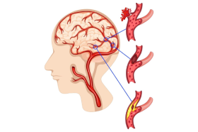
A migraine is not just a headache, it’s a neurological condition that can become disabling. They are often described as a throbbing or pressure like pain that typically affects one side of the head. Migraines are associated with nausea, vomiting, and sensitivity to light and sound. These episodes can negatively impact mood, concentration, and the ability to think clearly (brain fog). If left untreated, migraines can progressively worsen over time leading to longer, more frequent attacks per month. Inside our body, there is a protein called Calcitonin Gene-Related Peptide (CGRP) which activates pain receptors responsible for migraines. At Neurovascular centers we target the cause of migraine rather than mask the symptoms. Using a minimally invasive procedure, we embolize the artery responsible for the release of CGRP neuropeptide effectively eliminating migraine attacks. This procedure is performed as an outpatient with little to now down time.

An aneurysm is a weak area in a blood vessel that usually enlarges. It’s often described as a “ballooning” of the blood vessel that can cause bleeding in the brain called subarachnoid hemorrhage.
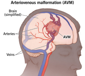
Arteriovenous malformation (AVM) is an abnormal connection between the arteries and veins that supply the brain. This tangle of blood vessels in the brain or on its surface can cause headaches, seizures, and brain hemorrhage.
A dural arteriovenous fistula (DAVF) is an abnormal connection between an artery and a vein within the covering of the brain protective surface (dura mater) called a fistula.
Similar to a arteriovenous fistula in the brain, spinal dural fistula are an abnormal connection between an artery and a vein located on the layer covering of the spinal cord.
.png)
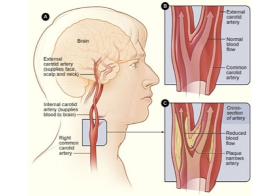
Like the arteries in the legs and heart, atherosclerosis is harding and narrowing of the blood vessels commonly can occur in the neck and brain vessels.
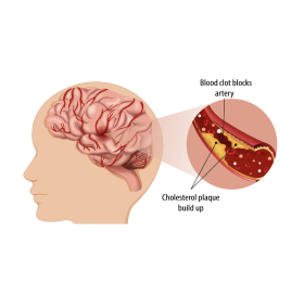
Intracranial atherosclerosis is harding and narrowing of the blood vessels in the arteries that supply blood to the brain. Similar to carotid artery atherosclerotic disease, atherosclerosis or harding of the blood vessles can also affect the arteries that supply blood to the brain.
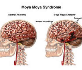
Moyamoya disease is a genetic disorder of the blood vessels in the brain that causes the arteries in the brain to become. narrow and eventually blocked. Often, tiny vessels called "moyamoya" vessels" form in the area of the narrowed or blocked vessels to compensate for the blockage.
Also known as idiopathic intracranial hypertension, is a disorder caused by elevated pressure on the brain which results in severe headaches, blurry vision, papilledema, and permanent blindness if not treated properly.
A migraine is not just a headache, it’s a neurological condition that can become disabling. They are often described as a throbbing or pressure like pain that typically affects one side of the head. Migraines are associated with nausea, vomiting, and sensitivity to light and sound. These episodes can negatively impact mood, concentration, and the ability to think clearly (brain fog). If left untreated, migraines can progressively worsen over time leading to longer, more frequent attacks per month. Inside our body, there is a protein called Calcitonin Gene-Related Peptide (CGRP) which activates pain receptors responsible for migraines. At Neurovascular centers we target the cause of migraine rather than mask the symptoms. Using a minimally invasive procedure, we embolize the artery responsible for the release of CGRP neuropeptide effectively eliminating migraine attacks. This procedure is performed as an outpatient with little to now down time.
An aneurysm is a weak area in a blood vessel that usually enlarges. It’s often described as a “ballooning” of the blood vessel that can cause bleeding in the brain called subarachnoid hemorrhage.
Subarachnoid hemorrhage (SAH) refers to bleeding within the subarachnoid space, which is the area between the brain and the tissues that cover the brain. Vascular Neurologist and a Endovascular Neurosurgeon are doctors trained to determine if you are at risk for SAH. Endovascular repair is the least invasive way to treat and prevent future rupture. Our main focus is to evaluate and decide if treatment is necessary.
Arteriovenous malformation (AVM) is an abnormal connection between the arteries and veins that supply the brain. This tangle of blood vessels in the brain or on its surface can cause headaches, seizures, and brain hemorrhage.
A dural arteriovenous fistula (DAVF) is an abnormal connection between an artery and a vein within the covering of the brain protective surface (dura mater) called a fistula.
Similar to a arteriovenous fistula in the brain, spinal dural fistula are an abnormal connection between an artery and a vein located on the layer covering of the spinal cord.
Like the arteries in the legs and heart, atherosclerosis is harding and narrowing of the blood vessels commonly can occur in the neck and brain vessels.
Intracranial atherosclerosis is harding and narrowing of the blood vessels in the arteries that supply blood to the brain. Similar to carotid artery atherosclerotic disease, atherosclerosis or harding of the blood vessles can also affect the arteries that supply blood to the brain.
Moyamoya disease is a genetic disorder of the blood vessels in the brain that causes the arteries in the brain to become. narrow and eventually blocked. Often, tiny vessels called "moyamoya" vessels" form in the area of the narrowed or blocked vessels to compensate for the blockage.
Also known as idiopathic intracranial hypertension, is a disorder caused by elevated pressure on the brain which results in severe headaches, blurry vision, papilledema, and permanent blindness if not treated properly.